
Page 8 - An ultrasound-driven immune-boosting molecular machine for systemic tumor suppression
P. 8
SCIENCE ADVANCES | RESEARCH ARTICLE
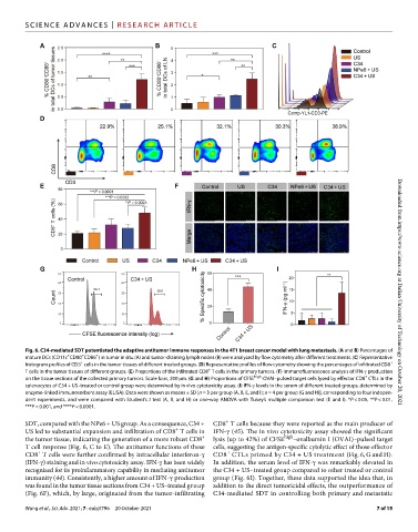
Fig. 6. C34-mediated SDT potentiated the adaptive antitumor immune responses in the 4T1 breast cancer model with lung metastasis. (A and B) Percentages of Downloaded from https://www.science.org at Dalian University of Technology on October 20, 2021
+
+
+
mature DCs (CD11c CD80 CD86 ) in tumor in situ (A) and tumor-draining lymph nodes (B) were analyzed by flow cytometry after different treatments. (C) Representative
+
+
histogram profiles of CD3 cells in the tumor tissues of different treated groups. (D) Representative profiles of flow cytometry showing the percentages of infiltrated CD8
+
T cells in the tumor tissues of different groups. (E) Proportions of the infiltrated CD8 T cells in the primary tumors. (F) Immunofluorescence analysis of IFN- production
+
on the tissue sections of the collected primary tumors. Scale bars, 200 m. (G and H) Proportions of CFSE high -OVAI–pulsed target cells lysed by effector CD8 CTLs in the
splenocytes of C34 + US–treated or control group were determined by in vivo cytotoxicity assay. (I) IFN- levels in the serum of different treated groups, determined by
enzyme-linked immunosorbent assay (ELISA). Data were shown as means ± SD [n = 3 per group (A, B, E, and I); n = 4 per group (G and H)], corresponding to four indepen-
dent experiments, and were compared with Student’s t test (A, B, and H) or one-way ANOVA with Tukey’s multiple comparison test (E and I). *P < 0.05, **P < 0.01,
***P < 0.001, and ****P < 0.0001.
+
SDT, compared with the NPe6 + US group. As a consequence, C34 + CD8 T cells because they were reported as the main producer of
+
US led to substantial expansion and infiltration of CD8 T cells in IFN- (45). The in vivo cytotoxicity assay showed the significant
+
the tumor tissue, indicating the generation of a more robust CD8 lysis (up to 42%) of CFSE high –ovalbumin I (OVAI)–pulsed target
T cell response (Fig. 6, C to E). The antitumor functions of these cells, suggesting the antigen-specific cytolytic effect of those effector
+
+
CD8 T cells were further confirmed by intracellular interferon- CD8 CTLs primed by C34 + US treatment (Fig. 6, G and H).
(IFN-) staining and in vivo cytotoxicity assay. IFN- has been widely In addition, the serum level of IFN- was remarkably elevated in
recognized for its proinflammatory capability in mediating antitumor the C34 + US–treated group compared to other treated or control
immunity (44). Consistently, a higher amount of IFN- production group (Fig. 6I). Together, these data supported the idea that, in
was found in the tumor tissue sections from C34 + US–treated group addition to the direct tumoricidal effects, the outperformance of
(Fig. 6F), which, by large, originated from the tumor-infiltrating C34-mediated SDT in controlling both primary and metastatic
Wang et al., Sci. Adv. 2021; 7 : eabj4796 20 October 2021 7 of 15