
Page 10 - An ultrasound-driven immune-boosting molecular machine for systemic tumor suppression
P. 10
SCIENCE ADVANCES | RESEARCH ARTICLE
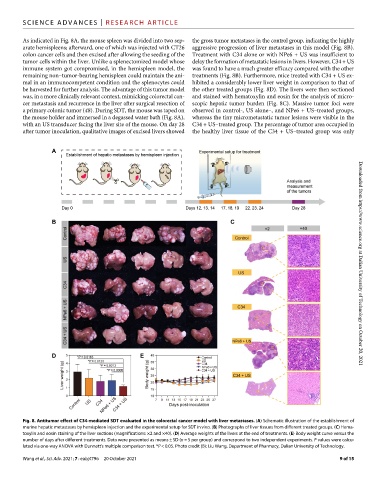
As indicated in Fig. 8A, the mouse spleen was divided into two sep- the gross tumor metastases in the control group, indicating the highly
arate hemispleens; afterward, one of which was injected with CT26 aggressive progression of liver metastases in this model (Fig. 8B).
colon cancer cells and then excised after allowing the seeding of the Treatment with C34 alone or with NPe6 + US was insufficient to
tumor cells within the liver. Unlike a splenectomized model whose delay the formation of metastatic lesions in livers. However, C34 + US
immune system got compromised, in the hemispleen model, the was found to have a much greater efficacy compared with the other
remaining non–tumor-bearing hemispleen could maintain the ani- treatments (Fig. 8B). Furthermore, mice treated with C34 + US ex-
mal in an immunocompetent condition and the splenocytes could hibited a considerably lower liver weight in comparison to that of
be harvested for further analysis. The advantage of this tumor model the other treated groups (Fig. 8D). The livers were then sectioned
was, in a more clinically relevant context, mimicking colorectal can- and stained with hematoxylin and eosin for the analysis of micro-
cer metastasis and recurrence in the liver after surgical resection of scopic hepatic tumor burden (Fig. 8C). Massive tumor foci were
a primary colonic tumor (48). During SDT, the mouse was taped on observed in control-, US alone–, and NPe6 + US–treated groups,
the mouse holder and immersed in a degassed water bath (Fig. 8A), whereas the tiny micrometastatic tumor lesions were visible in the
with an US transducer facing the liver site of the mouse. On day 28 C34 + US–treated group. The percentage of tumor area occupied in
after tumor inoculation, qualitative images of excised livers showed the healthy liver tissue of the C34 + US–treated group was only Downloaded from https://www.science.org at Dalian University of Technology on October 20, 2021
Fig. 8. Antitumor effect of C34-mediated SDT evaluated in the colorectal cancer model with liver metastases. (A) Schematic illustration of the establishment of
murine hepatic metastases by hemispleen injection and the experimental setup for SDT in vivo. (B) Photographs of liver tissues from different treated groups. (C) Hema-
toxylin and eosin staining of the liver sections (magnifications: ×2 and ×40). (D) Average weights of the livers at the end of treatments. (E) Body weight curve versus the
number of days after different treatments. Data were presented as means ± SD (n = 5 per group) and correspond to two independent experiments. P values were calcu-
lated via one-way ANOVA with Dunnett’s multiple comparison test. *P < 0.05. Photo credit (B): Liu Wang, Department of Pharmacy, Dalian University of Technology.
Wang et al., Sci. Adv. 2021; 7 : eabj4796 20 October 2021 9 of 15