
Page 4 - An ultrasound-driven immune-boosting molecular machine for systemic tumor suppression
P. 4
SCIENCE ADVANCES | RESEARCH ARTICLE
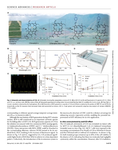
Fig. 2. Schematic and characterization of C34. (A) Schematic showing the preparation process of C34. (B and C) UV-vis (B) and fluorescence (C) spectra of CHC, NPe6, Downloaded from https://www.science.org at Dalian University of Technology on October 20, 2021
and C34. a.u., arbitrary units. (D) Illustration of the self-designed experimental configuration that provided the free-field US condition for in vitro study. (E) Free-field ul-
trasonic waveform detected by the hydrophone. (F and G) Detection of ROS generation capacity of C34 and NPe6 in solution by the probes of DPBF (F) and DCFH (G),
respectively, as the US exposure time increased. Data were presented as means ± SD (n = 3 per group) and compared using two-way analysis of variance (ANOVA).
****P < 0.0001.
corresponding to different spatial-average temporal-average inten- the macrocyclic structure of CHC could be a distinct advantage for
sity (IS ATA) is shown in table S1. enhancing acoustic responsive activity, enabling the potential im-
Although the mechanism of ROS generation during SDT remains provement of SDT efficiency for in vivo application.
unclear, ROS is still considered as an important cytotoxic species
for the killing effect of SDT (34). ROS generation capacity of C34 in In vitro sonocytotoxicity and ICD effect
the solution was assayed with two commercial ROS-sensing agents, The US-induced cytotoxicity of C34 was evaluated on tumor cells
1,3-diphenylisobenzofuran (DPBF) and 2′,7′-dichlorofluorescin by MTT [3-(4,5-dimethylthiazol-2-yl)-2,5-diphenyl tetrazolium
(DCFH). Upon interaction with ROS, DPBF could be bleached to bromide] assay in vitro (Fig. 3A). MCF-7 cells were exposed to the
the corresponding diketone, whereas DCFH turned to be its oxi- increasing concentrations (0 to 50 M) of C34 or NPe6 for 0.5 hours
dized form, DCF, resulting in the increase of fluorescent signal. As and then followed with or without US irradiation. As shown in fig.
shown in Fig. 2 (F and G), triggered by US, C34 produced signifi- S3, both treated groups remained up to 85% of the cell viability in
cantly higher levels of ROS than NPe6 in solution in a time-dependent the absence of US, indicating that these two molecules had negligible
pattern. Collectively, the introduction of the -extension system to cytotoxicity within the concentration. However, after US irradiation,
Wang et al., Sci. Adv. 2021; 7 : eabj4796 20 October 2021 3 of 15